Home › Unlabelled › Diagram Of Liver Gallbladder And Pancreas - Gall Bladder Pancreas
Diagram Of Liver Gallbladder And Pancreas - Gall Bladder Pancreas
Diagram Of Liver Gallbladder And Pancreas - Gall Bladder Pancreas. After you eat a meal, a substance called cholecystokinin is secreted by cells in the walls of the duodendum. The stomach forms part of the gastrointestinal tract between the esophagus and the duodenum (the first section of the small intestine). The duodenal cells release the hormone cholecystokinin d. The liver pancreas and gallbladder are considered accessory digestive organs but their roles in the digestive system are vital. Liver, stomach, pancreas, gallbladder and spleen detailed anatomy drawing on a white background liver functions.
Liver gallbladder stomach location in the human body. You may also develop yellowish eyes and skin which is called jaundice. A number of disorders can occur in the biliary system. The liver is divided into 8 segments based on its blood supply. Liver, gall bladder, and pancreas objectives:
Editable Liver Powerpoint Diagram Pslides from pslides.com In this image, you will find liver, gallbladder, stomach, duodenum in it. Figure 1.1 schematic diagram of the segmental anatomy of the liver. List two examples of polysaccharides. The bile ducts are a series of tubes that drain bile from the liver and either direct it to the gallbladder for temporary storage or pass it into the duodenum where it can be expelled with the feces. Liver gallbladder stomach location in the human body. Gallbladder is such an organ that stays hidden inside human body and in the minds of anatomists and doctors. Startradiology insulin is a hormone produced by the beta cells of the pancreatic islets of the pancreas. Our latest youtube film is ready to run.
Recognize liver, gall bladder, and pancreas.
Thus, couinaud described the left. A number of disorders can occur in the biliary system. The superior part of the duodenum, hepatic flexure and proximal transverse colon, are posteriorly related to it. The liver is divided into 8 segments based on its blood supply. Bile helps in absorption of digested fat in food. Liver, gall bladder, and pancreas objectives: A hollow muscular organ about the size of 2 closed fists, the stomach is located inferior to the diaphragm and lateral to the liver on the left side of the abdominal cavity. The pancreas and bile duct (biliary) systems together form an important part of the digestive system. As part of the digestive system, the gallbladder and pancreas help you break down food. The gallbladder is a small hollow intraperitoneal organ. The gallbladder wall lacks the thick muscular layers of the bowel wall, but still has a mucosa, lamina propria, smooth muscle, and serosa (except on hepatic surface). Draw a labelled diagram showing the interconnections between the liver, gall bladder, pancreas and small intestine. The head of the pancreas is on the right side of the abdomen and is connected to the duodenum (the.
Start studying liver and pancreas. The duodenal cells release the hormone cholecystokinin d. The gallbladder lies under the liver and frequently (70 %) invades the liver by direct extension. List two examples of polysaccharides. Startradiology insulin is a hormone produced by the beta cells of the pancreatic islets of the pancreas.
Structure Of Liver Gallbladder Pancreas And Duodenum Diagram Quizlet from o.quizlet.com The superior part of the duodenum, hepatic flexure and proximal transverse colon, are posteriorly related to it. The pancreas and bile duct (biliary) systems together form an important part of the digestive system. As part of the digestive system, the gallbladder and pancreas help you break down food. The pancreas has both an endocrine and a digestive exocrine function. Slide 194 liver, gall bladder h&e view virtual slide slide 195m liver, gall bladder masson view virtual slide upon gross examination of slides 194 and 195m (i.e. Our latest youtube film is ready to run. The hepatic artery carries blood from the aorta to the liver, whereas the portal vein carries blood containing the digested nutrients from the entire gastrointestinal tract, and also from. After you eat a meal, a substance called cholecystokinin is secreted by cells in the walls of the duodendum.
The gallbladder wall lacks the thick muscular layers of the bowel wall, but still has a mucosa, lamina propria, smooth muscle, and serosa (except on hepatic surface).
The liver pancreas and gallbladder are considered accessory digestive organs but their roles in the digestive system are vital. The liver produces bile continuously, but the body only needs it a few times a day. Start studying liver gallbladder and pancreas model. The fullest, most distal part of the gallbladder. Pancreas and liver location in the human body. Recognize liver, gall bladder, and pancreas. You may also develop yellowish eyes and skin which is called jaundice. National institute of diabetes and digestive and kidney diseases, national institutes of health. The hepatic artery carries blood from the aorta to the liver, whereas the portal vein carries blood containing the digested nutrients from the entire gastrointestinal tract, and also from. Its main function is to store bile that is produced from the liver. As part of the digestive system, the gallbladder and pancreas help you break down food. Thus, couinaud described the left. Our latest youtube film is ready to run.
The biliary system, including the liver, pancreas and gallbladder, form a part of the body's digestive system that is responsible for nutrient absorption and waste disposal. Anatomy of the stomach, gallbladder, and pancreas stomach. Liver, gall bladder, and pancreas objectives: One abnormal characteristic is the liver's regenerative abilities. The bile ducts are a series of tubes that drain bile from the liver and either direct it to the gallbladder for temporary storage or pass it into the duodenum where it can be expelled with the feces.
Liver Gall Bladder Bile Duct Pancreas Macs Clinic from macsforcancer.com The liver produces bile continuously, but the body only needs it a few times a day. Learn vocabulary, terms, and more with flashcards, games, and other study tools. The duodenal cells release the hormone cholecystokinin d. The gallbladder lies under the liver and frequently (70 %) invades the liver by direct extension. Gallbladder is such an organ that stays hidden inside human body and in the minds of anatomists and doctors. At the end of this laboratory you should be able to: Identify and understand the functional significance of the vascular supply and bile drainage of the liver. The fullest, most distal part of the gallbladder.
The pancreas has both an endocrine and a digestive exocrine function.
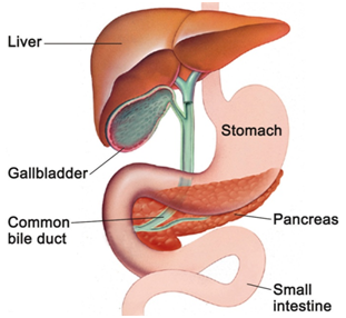
The liver is seen above the stomach, gall bladder, and pancreas. Slide 194 liver, gall bladder h&e view virtual slide slide 195m liver, gall bladder masson view virtual slide upon gross examination of slides 194 and 195m (i.e. Pancreas and liver location in the human body. Identify and understand the functional significance of the vascular supply and bile drainage of the liver. Figure 1.1 schematic diagram of the segmental anatomy of the liver. The pancreas and liver produce juices (pancreatic juice and bile) which help in the process of digestion (i.e. When autocomplete results are available use up and down arrows to review and enter to select. Pancreas and liver location in the human body. A hollow muscular organ about the size of 2 closed fists, the stomach is located inferior to the diaphragm and lateral to the liver on the left side of the abdominal cavity. The liver is connected to two large blood vessels, the hepatic artery and the portal vein. Liver, stomach, pancreas, gallbladder and spleen detailed anatomy drawing on a white background liver functions. The pancreas has both an endocrine and a digestive exocrine function. Liver gallbladder stomach location in the human body.
The gallbladder wall lacks the thick muscular layers of the bowel wall, but still has a mucosa, lamina propria, smooth muscle, and serosa (except on hepatic surface) diagram of liver. Learn vocabulary, terms, and more with flashcards, games, and other study tools.


comment 0 comments
more_vert